


Hip joint painIs specific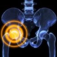 discomfort, discomfort due to pathology of the upper femur, tibia, adjacent soft tissue structures. In terms of intensity, they vary from weak to unbearable, in essence they can be smoldering, sharp, pressing, stinging, exploding, drilling, etc. Usually they depend on the load, time of day and other conditions. another factor. The cause of the pain is determined using X-rays, CT, MRI, ultrasound, arthroscopy, and other studies. Pain medication and limb rest are recommended until a diagnosis is made.
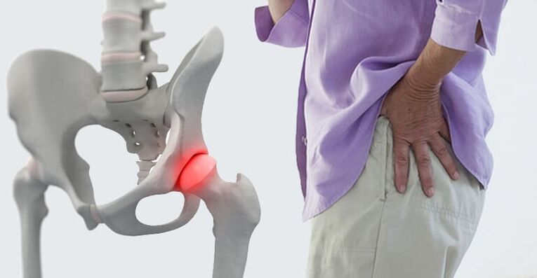
Causes of hip pain
Soft tissue injury
The most common cause of traumatic pain is a stiff hip. Occurs with a fall or a direct impact, manifested by acute moderate pain, rapidly becoming dull, diminishing and disappearing within a few days, in severe cases - weeks. The supports are preserved, the movements are a bit limited. Edema is detected locally, possibly with bruising.
Injury to the ligaments of the hip joint is very rare, usually as a result of road traffic accidents and sports injuries, accompanied by severe pain, sometimes - a feeling of cracking (such as tissue tearing). The pain subsides somewhat, then usually increases again due to edema. Swelling from the joint extending to the groin and thigh area.
The degree of dysfunction in injury to the ligamentous apparatus depends on the severity of the injury (stretch, tear, rupture), ranging from mild limitation to inability to support the leg. The pain increases with displacement, movement in the opposite direction of the injured ligament.
Osteoarthritis
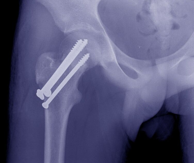
Hip fractures often occur in older adults as a result of domestic or street trauma. A characteristic feature, especially in the presence of osteoporosis, is the absence of a syndrome of severe pain, mild edema. At rest, the pain is deep, dull, moderate or insignificant; when moving, the pain is strongly increased. Support is sometimes withheld. A common symptom is an inability to lift the leg straight from a prone position (symptom of a stuck heel).
Transverse fractures are more commonly diagnosed in middle-aged and young adults and develop as a result of high-energy trauma. Unlike ankle fractures, they are accompanied by unbearable sharp radiating pain. Then the pain subsided, but was still very strong, uncomfortable. Joints are swollen and bruised. Movement is severely limited. Support is not possible.
Isolated fractures in older people are very rare; they are found in children and young adults; They are formed by a fall, direct impact, or strong muscle contraction. Acute, very severe pain, localized mainly on the outer surface of the joint. Due to the increased pain, the patient avoids vigorous exercise.
Dislocations of the hip occur from falls from heights, road traffic and industrial injuries, presenting with unbearable sharp pain that barely subsides until relieved. The joint is deformed, the leg is shortened, bent at the knee joint, turned outward, less inward (depending on the type of dislocation). Lifting and moving is not possible, when trying to move, the resistance of the spring is determined.
Tubular fractures develop singly or in association with hip dislocation. Characterized by acute explosive pain in the depth of the hip joint. After that, the pain subsided somewhat, but remained intense, hindering any movement. Legs shortened, turned outward. Support is not possible.
Degenerative process
With coxarthrosis in the early stages, the pain is cyclical, dull, uncertain locality, appearing at the end of the day or after a significant load, sometimes spreading to the hip, knee joints. There may be a mild sensation of stiffness, which quickly passes with the onset of movement. Then, the intensity of pain increases, pain is noted not only with movement but also at rest. After exertion, the patient begins to limp. Movement is somewhat limited.
In severe coxarthrosis, the pain is deep, diffuse, constant, aching, twisting. Disturbing both day and night. The ability to cope with stress decreases, when walking, the patient has to use a cane. Movement is significantly limited, the affected leg is shortened resulting in increased load on the joint, and pain increases when walking and standing.
Inflammatory disease of the hip joint in its course resembles subacute arthritis. The pain is moderate, diffuse, transient, associated with a crunching sound, with limited range of motion. When the organs in the joint are compromised, obstruction occurs, characterized by severe pain, inability or significant limitation of movement. After cessation of the arthropod invasion, the listed symptoms should disappear.
Plantar fasciitis is usually formed with arthritis of the hip, accompanied by inflammatory degenerative damage of the tendons of the gluteal muscle at the point of attachment to the greater gluteal muscle, manifested by pain in the affected area when lying down. upside down. position on the affected side. Pain increases when trying to flex the hip with resistance.
Bone nutritional disorders
Perthes disease develops in children and adolescents and is characterized by partial necrosis of the femoral head, initially accompanied by a dull, non-intense deep pain, sometimes radiating to the knee and hip. After a few months, the pain increases sharply, becomes constant, sharp pain, fatigue. Joints swell, movement is limited, and a limp appears. Then the pain subsides, the degree of recovery of joint functions varies.
Aseptic necrosis of the femoral head resembles Perthes disease, but it is detected in adults, progressing less favorably, in half of the cases bilateral. At first, the pain is cyclical in nature. Then the pain syndrome gradually increases, appearing at night. At the peak of clinical manifestations is severe pain that makes the patient completely lose the ability to lean on the leg. Then the pain subsided. Limited range of motion progressed over about 2 years, resulting in hip sclerosis, spasticity, and shortening of the extremities.
Solitary bony cyst that forms in the proximal part of the thigh in 10 to 15 year old boys, accompanied by intermittent, non-severe pain in the hip. Edema is usually absent, with prolonged contractures often developing, especially in young children. Because of the mild symptoms, the cause of treatment is pathological fracture or increasing limitation of movement.
Arthritis
Aseptic arthritis is manifested by wave-like pain in the joints, which increases in the hours before morning. The degree of pain varies from insignificant to acute, strong, continuous, with significant limitation of physical activity. Stiffness, swelling, redness and local elevation of temperature were noted. Feel pain.
In rheumatoid arthritis, the hip joint is rarely ankylosing, the damage is symmetrical in nature. Cyclic pain first appears on the basis of the change of seasons (autumn, spring), pronounced changes in weather conditions, during the period of hormonal changes after childbirth or during menopause. Moderate or weak pain, diffuse, pulling or aching, intensifying to palpation. It is associated with recurrent synovitis, edema, congestion, hyperthermia, increased mobility limitation.
Infectious arthritis develops with hematogenous or lymphoid spread, less often - with the entry of pathogens into the joint from adjacent tissues. Usually acute onset with rapidly increasing pain. Severe pain, convulsions, lacrimation, rupture, discomfort at rest, aggravated by movement, so the limb must be kept in a forced position. The patient has fever, chills, sweating, severe weakness, edema, joint redness, and increased local temperature.
If not treated promptly, bacterial arthritis can turn into hip arthritis - a purulent inflammation of all tissues of the hip joint. It is characterized by a severe course with very acute widespread throbbing pains, fever, severe weakness, presyncope, marked hyperemia, and hyperthermia.
Other inflammatory diseases
Osteoarthritis of the upper femur can be caused by a hematoma, after trauma, or after surgery. Osteomyelitis presents with localized, very acute pain, convulsions, lacrimation or dull pain, so the patient avoids even the slightest limb movements. There is a marked increase in body temperature, severe intoxication.
Posttraumatic and postoperative osteomyelitis occurs with similar, but less pronounced, symptoms. Usually, with a more gradual onset against the background of an open fracture or postoperative wound, pus appears. Hip pain gradually increased over 1-2 weeks in parallel with the development of local inflammatory markers.
Bursitis develops against the background of trauma, other diseases of the hip joint, it rarely becomes a manifestation of an allergy. In acute bursitis, the pain is usually small, dull, flared, and gradually increased due to increased fluid in the joint. The joint is swollen, slightly painful to the touch, and the symptoms are determined to be fluctuating. Chronic bursitis is asymptomatic, accompanied by pain and weakness.
With episodic hydrocephalus, the pain is also insignificant, accompanied by discomfort, limitation of movement, and disappears within 3-5 days after the effusion is reabsorbed. They renew at regular intervals, individually for each patient, due to the repeated accumulation of fluid in the joints.
Specific infection
Tuberculosis of the hip is a common form of tuberculosis of the joints, manifested by general weakness, fatigue, and paralysis. Then there are weak or painful contractions of the muscles, a transient pain in the joints when walking. The patient begins to spare limbs. As the pain progresses, they become moderate, diffuse, radiating to the knee, complemented by swelling, redness, and bursitis. A development protection contract.
Joint pain, including the hip, may be present with brucellosis. In acute and subacute forms, painful sensations of pulling, twisting, associated with periodic fever, lymphadenopathy, skin rash. In a chronic process, the pain syndrome resembles that in aseptic arthritis, deformities are formed over time.
Birth defects
Manifestations of hip dysplasia are determined by the degree of abnormality of the femoral head and tibia. With congenital complete dislocation, pain occurs soon after the child begins to walk, accompanied by a limp. With a moderate degree of myopia, painful sensations occur at the age of 5-6 years, which is directly related to the load on the legs.
With a mild stage, the pathology is asymptomatic for a long time, the pain syndrome manifests itself with the development of dysplastic coxarthrosis at the age of 25-30 years. The telltale signs of such joint disease are rapidly increasing pain, early onset of pain at rest and at night, and increasing limitation of movement. All forms of dysplasia are accompanied by asymmetry of skin folds, "click" symptoms, and limited mobility. In case of dislocation, note shortening of the limb.
Neoplasms
For benign neoplasms, a typical course is asymptomatic. The pain is mild, intermittent, and usually does not progress for many years. Tumor growth is accompanied by a slowly increasing pain syndrome, recurrent synovitis. In the hip joint area, osteosarcoma, osteosarcoma, osteoblastoma, choroidal tumor are more often detected.
Malignant neoplasms (osteoma, choroidal neoplasm) are characterized by rapid progression of pain syndrome and other pathological manifestations. At first, the pain is mild, short-term, not specific, sometimes worse at night. Then they become sharp, permanent, cut, envelop, spread to the entire joint. Vaginal area is swollen and deformed. Weight loss, weakness, and ill health are noted. With advanced cancer, pain and discomfort can only be eliminated with anesthesia.
Other reasons
Pain in the hip is sometimes accompanied by dorsal plexitis and sciatic nerve pain, however, they often occupy a minor role in the clinical picture of the disease, fading in comparison to the severe pain inback buttock and thighs, weak limbs. and sensitivity disorders.
This focal pain syndrome is often found in osteonecrosis and disc herniation. Pain can be detected with spondylitis, degenerative spondylolisthesis, and scoliosis. Dull, cyclical, aching pains, often intensifying during the exacerbation of the underlying disease. The cause of their appearance can be a constant overload of the joints or the development of coxarthrosis.
Sometimes joint pain is triggered by mental illness or a depressive disorder. Diabetes mellitus is often accompanied by intussusception, balanitis, and other lesions of the peristaltic soft tissues. Drug-induced arthropathy can occur while taking certain medications.
Diagnose
In the case of trauma, diagnostic measures are carried out by traumatologists. Degenerative and inflammatory diseases managed by chiropractors and rheumatologists. In the case of purulent processes, the involvement of a surgeon is necessary. The examination includes collection of complaints, medical history, physical examination, and additional research. Taking into account the peculiarities of the pathological process, the following methods can be used:
- X-ray. It is the basic technique for most joint diseases. Detection of fractures, dislocations, changes in the contour of the femoral and femur, margin and intraosseous defects, bony growth, and narrowing of joint spaces
- Supersonic.Most informative when studying soft tissues. Reveal signs of inflammatory and degenerative processes, calcified areas. Used to diagnose joint effusion, cramps.
- MRI and CT.Clarification techniques are used where data are not clear from primary studies, to clarify the nature, prevalence, and location of the pathological focus. Can be done with contrast.
- Joint perforation.Of a diagnostic or therapeutic and diagnostic nature. Allows you to eliminate effusion, study the composition of intra-articular fluid, determine the causative agent of infection by laboratory tests.
- Arthroscopy.During the visual examination of the joint, the doctor will assess the condition of the bony structures and soft tissues, if necessary, take a biopsy sample for further histological examination and perform therapeutic measures.
- Laboratory tests.They are prescribed to identify inflammatory markers and signs of rheumatic diseases, to evaluate the general condition of the body, the functioning of various organs in serious infectious or systemic pathologies.
Treatment
Help before diagnosis
In case of severe injury, it is necessary to immobilize the joint by splinting from the foot to the armpit. In the case of a minor injury, simply resting the leg is enough. Cold is applied to the affected area. For severe pain, pain medication will be given. You can not perform active movements with limbs, leg loads. It is strictly forbidden to attempt to eliminate displacement or displacement of the bone.
Strategies for non-traumatic diseases are determined by symptoms. With mild symptoms, it is possible to ensure that the limb is rested, using local remedies with analgesic and anti-inflammatory effects. In case the child has fever, weakness, severe pain, edema and rapid rise in blood pressure, immediate medical attention should be sought.
Conservative therapy
Dislocation is a sign for immediate relief. In the case of fractures, bone traction is usually applied, after which the patient is operated on or immobilized with a plaster cast after calluses appear. In elderly patients with hip fractures, immobilization is allowed with a shock-reducing warm-up, which prevents rotation of the joint.
The remaining patients were advised to have hip arthroplasty. As indicated, orthopedic braces or additional devices (crutches, canes) should be used. Prescribing massage, physiotherapy exercises, physiotherapeutic procedures:
- laser therapy;
- acupuncture therapy;
- UHF;
- supersonic;
- drug electrophoresis;
- UHT.
It is possible to use NSAIDs, chondroprotectors, antibacterial drugs. Local agents are widely used. According to indications, arthrocentesis, intra- and peri-articular blockade with hormones, intra-articular injection of chondroprotectors, synovial fluid substitutes are performed.
Surgery
Operations on the hip are performed using an open approach and with the assistance of arthroscopic equipment. Taking into account the type of pathology, the following can be done:
- Traumatic injury:Open hip dislocation relief, glial reconstruction, cervical bone formation, trochanteric fracture.
- Degenerative process:Arthroscopy, arthroscopy, free tissue removal in joints.
- Tumor:Tumor removal, osteotomy, hip resorption, Io-abdominal amputation, Io-abdominal amputation.
In the case of spasticity, ankylosing spondylitis, scarring of the periarticular tissues, resurfacing surgery, arthroplasty, and arthroplasty can be performed. Internal medicine is an effective way to restore limb functions in diseases of various origins, accompanied by limitation of movement or destruction of joints.